Deep radiogenomics - classification of brain tumor to molecular subtypes based on magnetic resonance images
Radiogenomics is a challenging task of distinguishing molecular subtypes of a lesion based on radiological imaging data. The genomic type of a tumor is a valuable information and can help in guiding patient’s treatment.
The dataset used for development was obtained from The Cancer Imaging Archive (TCIA) and involved 110 cases of lower-grade glioma patients. Registers brain MR images with manual FLAIR abnormality segmentation masks are published as a Kaggle Dataset lgg-mri-segmentation.


Figure 1. Randomly sampled patches of tumors extracted from FLAIR sequences of brain MRI masked with manual segmentations. Each row corresponds to a separate genomic subtype cluster.
A method that we applied was transfer learning from a different brain MRI dataset containing scans from cases with tumors of a similar type. Obtained results show a notable association between imaging and genomic data. This provides strong evidence for genomic subtypes being exposed in MRI.
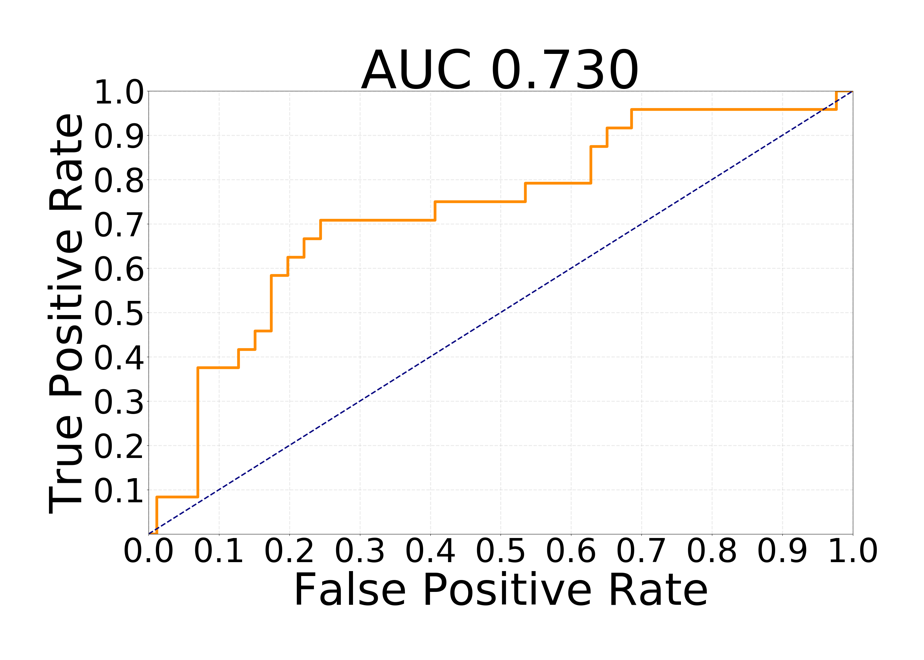
Figure 2. Receiver operating characteristic curve for the task of discriminating betwen tumor genomic subtypes of significantly different survivals times.
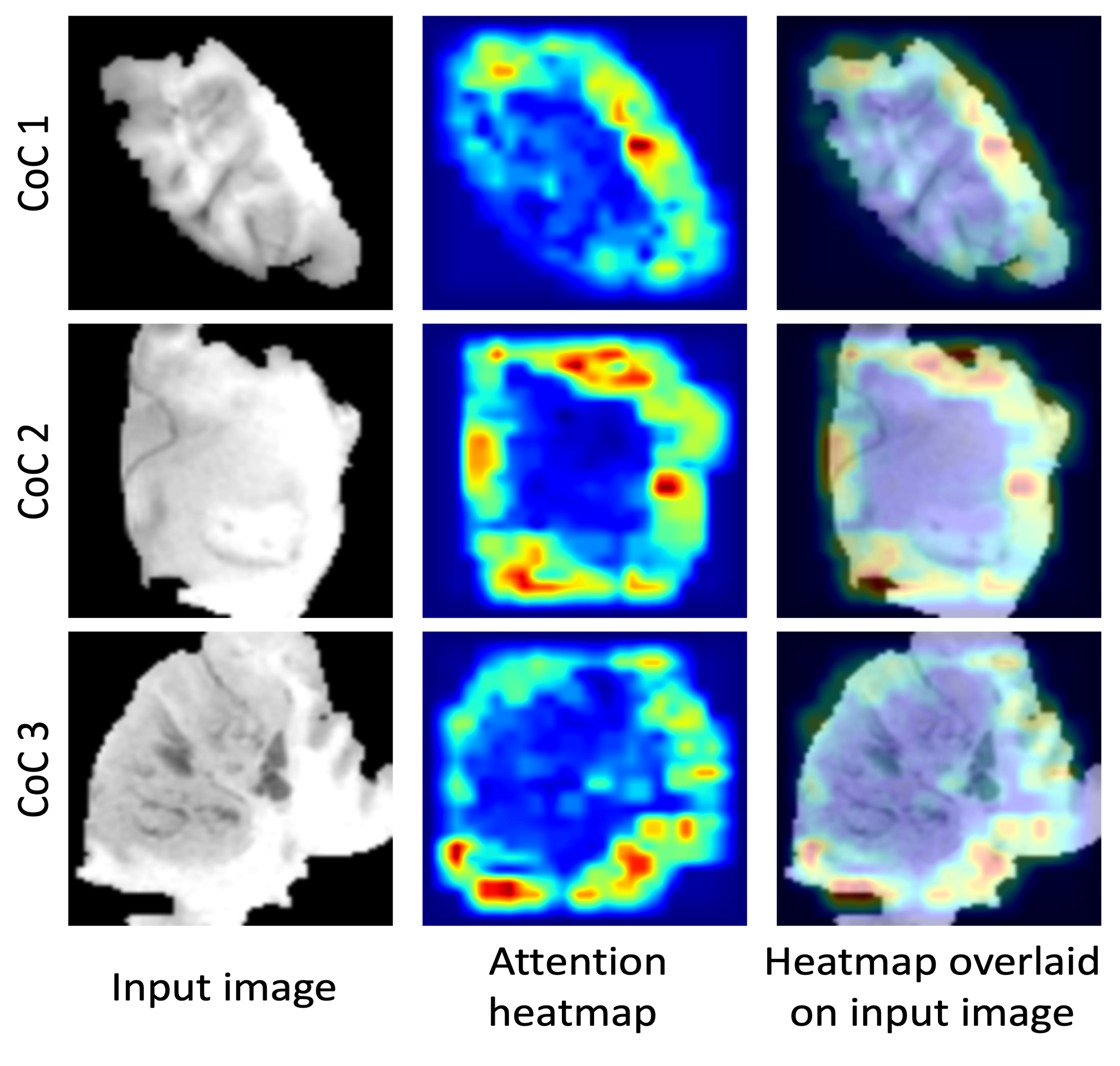
In Figure 3, we show network attention heatmaps, which indicate parts of the image responsible for prediction. Increased response by the network was for tumor margin regions of high irregularity.
While deep learning cannot yet replace genomic testing, it shows promise in aiding clinical decisions.
Links
- Full paper: Radiology: Artificial Intelligence
- Data: lgg-mri-segmentation